
Sensitivity vs Specificity

When developing diagnostic tests or evaluating results, it is important to understand how reliable those tests and therefore the results you are obtaining are. By using samples of known disease status, values such as sensitivity and specificity can be calculated that allow you to evaluate just that.

What do sensitivity values tell you?


The sensitivity of a test is also called the true positive rate (TPR) and is the proportion of samples that are genuinely positive that give a positive result using the test in question. For example, a test that correctly identifies all positive samples in a panel is very sensitive. Another test that only detects 60 % of the positive samples in the panel would be deemed to have lower sensitivity as it is missing positives and giving higher a false negative rate (FNR). Also referred to as type II errors, false negatives are the failure to reject a false null hypothesis (the null hypothesis being that the sample is negative).

What do specificity measures tell you?


The specificity of a test, also referred to as the true negative rate (TNR), is the proportion of samples that test negative using the test in question that are genuinely negative. For example, a test that identifies all healthy people as being negative for a particular illness is very specific. Another test that incorrectly identifies 30 % of healthy people as having the condition would be deemed to be less specific, having a higher false positive rate (FPR). Also referred to as type I errors, false positives are the rejection of a true null hypothesis (the null hypothesis being that the sample is negative).

Sensitivity vs specificity mnemonic


SnNouts and SpPins is a mnemonic to help you remember the difference between sensitivity and specificity.

SnNout: A test with a high sensitivity value (Sn) that, when negative (N), helps to rule out a disease (out).

SpPin: A test with a high specificity value (Sp) that, when positive (P) helps to rule in a disease (in).

How do I calculate sensitivity and specificity values?


An ideal test rarely overlooks the thing you are looking for (i.e., it is sensitive) and rarely mistakes it for something else (i.e. it is specific). Therefore, when evaluating diagnostic tests, it is important to calculate the sensitivity and specificity for that test to determine its effectiveness.

The sensitivity of a diagnostic test is expressed as the probability (as a percentage) that a sample tests positive given that the patient has the disease.

The following equation is used to calculate a test’s sensitivity:

Sensitivity = Number of true positives (Number of true positives + Number of false negatives)

           = Number of true positives Total number of individuals with the illness

The specificity of a test is expressed as the probability (as a percentage) that a test returns a negative result given that the that patient does not have the disease.

The following equation is used to calculate a test’s specificity:

Specificity =     Number of true negatives
(Number of true negatives + number of false positives)

           =     Number of true negatives Total number of individuals without the illness

Sensitivity vs specificity example


You have a new diagnostic test that you want to evaluate. You have a panel of validation samples where you know for certain whether they are definitely from diseased or healthy individuals for the condition you are testing for. Your sample panel consists of 150 positives and 400 negatives.

After running the samples through the assay, you compare your results to their known disease status and find:

True positives (test result positive and is genuinely positive) = 144

False positive (test result positive but is actually negative) = 12

True negatives (test result negative and is genuinely negative) = 388

False negative (test result negative but is actually positive) = 6

Sensitivity vs specificity table


Or, displayed in a contingency table:


Genuinely PositiveGenuinely NegativeRow Total
Test Positive14412156
Test Negative6388394
Column Total150400550

Sensitivity = 144 / (144 + 6)
= 144 / 150
= 0.96
= 96 % sensitive
Specificity = 388 / (388 + 12)
= 388 / 400
= 0.97
= 97 % specific

Are sensitivity and specificity the same as the positive predictive value (PPV) and negative predictive value (NPV)?


In short, no, although they are related. The positive predictive value (PPV) is the probability that a subject/sample that returns a positive result really is positive. The negative predictive value (NPV) is the probability that a subject/sample that returns a negative result really is negative. This sort of information can be very useful for discussing results with a patient for example, evaluating the reliability of any test they may have had. The same values used to calculate the sensitivity and specificity are also used to calculate the positive and negative predictive values. One way to look at it is that the sensitivity and specificity evaluate the test, whereas the PPV and NPV evaluate the results.

The positive predictive value is calculated using the following equation:

PPV = Number of true positives
(Number of true positives + Number of false positives)

       = Number of true positives     Number of samples that tested positive

The negative predictive value is calculated using the following equation:

NPV = Number of true negatives
(Number of true negatives + Number of false negatives)

       = Number of true negatives Number of samples that tested negative

Using the values from the example above:

PPV = 144 / (144 + 12)
        = 144 / 156
        = 0.923076923… = 92 %

NPV = 388 / (388 + 6)
        = 388 / 394
        = 0.984771573… = 98 %

So, if a test result is positive, there is a 92 % chance it is correct, if it is negative there is a 98 % chance it is correct.

The complementary value to the PPV is the false discovery rate (FDR), the complementary value of the NPV is the false omission rate (FOR) and equates to 1 minus the PPV or NPV respectively. The FDR is the proportion of results or “discoveries” that are false. The FOR is the proportion of false negatives which are incorrectly rejected. Essentially, the higher the PPV and NPV are, the lower the FDR and FOR will be - which is good news for the reliability of your test results.

How should I balance sensitivity with specificity? 


Where results are given on a sliding scale of values, rather than a definitive positive or negative, sensitivity and specificity values are especially important. They allow you to determine where to draw cut-offs for calling a result positive or negative, or maybe even suggest a grey area where a retest would be recommended. For example, by putting the cutoff for a positive result at a very low level (purple dashed line), you may capture all positive samples, and so the test is very sensitive. However, this may mean many samples that are actually negative could be regarded as positive, and so the test would be deemed to have poor specificity. Finding a balance is therefore vital for an effective and usable test.

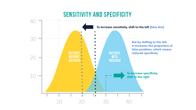
Using a receiver operating characteristic (ROC) curve can help to hit that sweet spot and balance false negatives with false positives. However, the context is also important as to whether false negatives are less problematic than false positives, or vice versa. For example, if it is imperative that all positives are identified – for example, in a matter of life and death, then you may be willing to tolerate a higher number of false positives to avoid missing any. Here, false positives can be screened out further down the line.


What is a ROC curve?


A ROC curve is a graphical representation showing how the sensitivity and specificity of a test vary in relation to one another. To construct a ROC curve, samples known to be positive or negative are measured using the test.

The TPR (sensitivity) is plotted against the FPR (1 - specificity) for given cut-off values to give a plot similar to the one below. Ideally a point around the shoulder of the curve is picked which both limits false positives whilst maximizing true positives.

A test that gave a ROC curve such as the yellow line would be no better than random guessing, pale blue is good, but a test represented by the dark blue line would be excellent. It would make cutoff determination relatively simple and yield a high true positive rate at very low false positives rate – sensitive and specific.

Source: Sensitivity vs Specificity

CÁC BÀI ĐĂNG ĐƯỢC XEM NHIỀU

Atlas CÁC DÒNG TẾ BÀO MÁU BÌNH THƯỜNG

ATLAS CÁC DÒNG TẾ BÀO MÁU BÌNH THƯỜNG DÒNG BẠCH CẦU   1. Hemocytoblast (Nguyên bào máu) 2. Myeloblast (Nguyên tủy bào) 3.Neutrophil promyelocyte (Tiền tủy bào trung tính) 4. Neutrophil myelocyte (Tủy bào trung tính) 5. Neutrophil metamyelocyte (Hậu tủy bào trung tính) 6.Neutrophil band (Bạch cầu đũa) 7. Neutrophil segmented (Bạch cầu đoạn trung tính) 8.  Neutrophil myelocyte/metamyelocyte/band/segmented (Tủy bào/Hậu tủy bào/bạch cầu đũa/bạch cầu đoạn trung tính)   9. Eosinophil promyelocyte (Tiền tủy bào ưa acid) 10. Eosinophil myelocyte (Tủy bào ưa acid) 11. Eosinophil metamyelocyte (Hậu tủy bào ưa acid) 12. Eosinophil band (Bạch cầu đũa ưa acid) 13. Eosinophil segmented (Bạch cầu đoạn ưa acid) 14. Neutrophil/Eosinophil segmented (Bạch cầu đoạn trung tính/Bạch cầu đoạn ưa acid) 15. Basophil myelocyte (Tủy bào ưa base) 16. Basophil segmented (Bạch cầu đoạn ưa base) DÒNG LYMPHO  17. Lymphoblast

CHỈ SỐ HỒNG CẦU LƯỚI THỰC (Corrected Reticulocyte Count - CRC) LÀ GÌ? TẠI SAO VIỆC XÁC ĐỊNH NÓ CÓ VAI TRÒ CỰC KỲ QUAN TRỌNG TRONG ĐÁNH GIÁ TÌNH TRẠNG THIẾU MÁU

CHỈ SỐ HỒNG CẦU LƯỚI THỰC (Corrected Reticulocyte Count - CRC) LÀ GÌ? TẠI SAO VIỆC XÁC ĐỊNH NÓ CÓ VAI TRÒ CỰC KỲ QUAN TRỌNG TRONG ĐÁNH GIÁ TÌNH TRẠNG THIẾU MÁU Hầu như các bạn đều biết đến Chỉ số Hồng cầu lưới máu ngoại vi (Reticulocyte-Ret) và vai trò quan trọng của nó. Tuy nhiên, việc đánh giá thiếu máu dựa vào chỉ số Ret có thể dẫn tới sai lầm, vậy tại sao lại sai lầm, và để tránh sai lầm trong đánh giá người ta dùng chỉ số gì? Câu trả lời, đó là CRC - chỉ số hồng cầu lưới thực. 1. Hồng cầu lưới ở máu ngoại vi (Ret) là gì? Vai trò của hồng cầu lưới 2. Sai lầm khi sử dụng chỉ số Ret trong đánh giá thiếu máu 3. Chỉ số hồng cầu lưới thực (CRC - Corrected reticulocyte count) 1. RETICULOCYTE COUNT (Chỉ số hồng cầu lưới máu ngoại vi) Hồng cầu lưới (RET) là các hồng cầu non được giải phóng từ tủy xương ra máu ngoại vi. Sau 24h ở máu ngoại vi, Ret sẽ "chín" và trở thành hồng cầu trưởng thành. CÁCH XÁC ĐỊNH Có thể dễ dàng xác định Ret bằng cách

TẠI SAO PHỤ NỮ MANG THAI LẠI CÓ SỰ TĂNG SỐ LƯỢNG BẠCH CẦU?

 TẠI SAO PHỤ NỮ MANG THAI LẠI CÓ SỰ TĂNG SỐ LƯỢNG BẠCH CẦU? Trong thời kỳ mang thai, số lượng bạch cầu tăng thêm kho ảng < 6 G/L. Tăng bạch cầu này xảy ra là do phản ứng stress sinh lý (the physiologic stress) gây ra bởi tình trạng mang thai.  (Stress sinh lý là một phản ứng của cơ thế đến tác nhân gây stress, ví dụ như sự thay đổi môi trường, hay một tác nhân kích thích, ở đây là tình trạng mang thai của cơ thể người nữ). Tăng bạch cầu đoạn trung tính (Neutrophils) là chủ yếu. Điều này có thể do sự suy giảm bạch cầu đoạn trung tính trong chương trình chết tế bào bạch cầu đoạn trung tính (neutrophilic apoptosis) khi mang thai. (Apoptosis hay sự chết tế bào theo chương trình là một quá trình xuyên suốt cuộc sống, giúp cơ thể loại bỏ các tế bào già cỗi, các tế bào không còn cần thiết, các tế bào sai hỏng, bị tổn thương có thể dẫn tới ung thư) Trong bào tương bạch cầu đoạn trung tính có các hạt đặc hiệu trung tính giúp hóa ứng động bạch cầu và thực bào tác nhân l

NHỮNG TÓM TẮT QUAN TRỌNG VỀ HỒNG CẦU LƯỚI VÀ CÁCH SỬ DỤNG CHỈ SỐ HỒNG CẦU LƯỚI

HỒNG CẦU LƯỚI (Reticulocytes and reticulocyte count) (Trong bài này CÓ NHIỀU KIẾN THỨC MỚI mà ít sách ở Việt Nam đề cập) 1. Sự quan trọng của hồng cầu lưới (Reticulocytes-Ret) Ret là các hồng cầu non mới được giải phóng từ tuỷ xương ra máu ngoại vi Sự xuất hiện Ret ở máu ngoại vi, là chỉ điểm (marker) cho thấy quá trình tạo hồng cầu có hiệu quả. Sự tạo hồng cầu có hiệu quả cho thấy, tuỷ xương phản ứng tốt với tình trạng thiếu máu. - Có mối tương quan giữa tăng tổng hợp và giải phóng Ret từ tuỷ xương ra máu ngoại vi, khi có tình trạng thiếu máu. 2. Có thể dễ dàng xác định được Ret ở máu ngoại vi bằng cách nhuộm máu tươi với thuốc nhuộm xanh methylene (hoặc có thể dùng xanh cresyl). Đặc điểm Ret sau nhuộm: Có những sợi ARN mảnh như sợi chỉ, nằm trong bào tương của các hồng cầu non 3. Sau 24 giờ ở máu ngoại vi, hồng cầu lưới sẽ "chín" và trở thành hồng cầu trưởng thành. Sự trưởng thành xảy ra được là nhờ sự giúp đỡ của đại thực bào ở lách. 4. Số

Tại sao thường sử dụng chống đông EDTA trong xét nghiệm HbA1c? Và có thể sử dụng chống đông khác (Heparin, NaF, Natri Citrat) được không?

Tại sao thường sử dụng ống chống đông EDTA để thu thập bênh phẩm máu thực hiện xét nghiệm HbA1c? Có thể sử dụng ống chống đông khác như (Na-Citrate , Heparin, Na-flouride) thay thế được không? Để trả lời cho câu hỏi này, tôi sẽ viện dẫn một nghiên cứu của Mailankot và các cộng sự (Mailankot M, Thomas T, Praveena P, Jacob J, Benjamin JR, Vasudevan DM, et al. Various anticoagulants and fluoride do not affect HbA1C level. Ind J Clin Biochem. 2012;27:209) Nghiên cứu : Tiến hành thu thập mẫu máu vào các ống chống đông EDTA, Heparin, Na-citrate, Na-flouride trên cùng một mẫu máu, rồi định lượng nồng độ (%) HbA1C trong 7 ngày, với cùng nhiệt độ bào quản 4 độ C. Kết quả cho thấy: KHÔNG CÓ SỰ THAY ĐỔI ĐÁNG KỂ NỒNG ĐỘ HbA1c ở các ống chống đông EDTA, Heparin, Na-citrate, Na-flouride khi bảo quản ở 4 độ C trong 7 ngày (xem hình ảnh biểu diễn kết quả bên dưới) Bảng thể hiện nồng độ HbA1C ở các mẫu có ĐTĐ và không ĐTĐ ở các ống chống đông khác nhau Đường biểu diễn nồng độ HbA1C ở n

Atlas TẾ BÀO MÁU TRONG BỆNH BẠCH CẦU LEUKEMIA

ATLAS TẾ BÀO MÁU TRONG BỆNH BẠCH CẦU LEUKEMIA 1. Acute Lymphatic Leukemia (ALL-L1) - Bạch cầu cấp dòng lympho thể L1 2.  Acute Lymphatic Leukemia (ALL-L2) - Bạch cầu cấp dòng lympho thể L2 3.  Acute Lymphatic Leukemia (ALL-L3) - Bạch cầu cấp dòng lympho thể L3 4.  Acute Myeloid Leukemia (AML-M0) - Bạch cầu cấp dòng tủy có độ biệt hóa tối thiểu 5.  Acute Myeloid Leukemia (AML-M1) - Bạch cầu cấp dòng tủy không trưởng thành 6.  Acute Myeloid Leukemia (AML-M2) - Bạch cầu cấp dòng tủy trưởng thành 7.  Acute Myeloid Leukemia (AML-M3) - Bạch cầu cấp thể tiền tủy bào 8.  Acute Myeloid Leukemia (AML-M3) Hypogranular - Bạch cầu cấp thể tiền tủy bào thể giảm hạt 9.  Acute Myeloid Leukemia (AML-M4) - Bạch cầu cấp dòng tủy và dòng mono 10. ACUTE MYELOID LEUKEMIA (AML-M5) - BẠCH CẦU CẤP DÒNG MONO 11. ACUTE MY

XÉT NGHIỆM YẾU TỐ RF (Rheumatoid Arthritis Factor) - XÉT NGHIỆM QUAN TRỌNG TRONG CHẨN ĐOÁN VIÊM KHỚP DẠNG THẤP

XÉT NGHIỆM YẾU TỐ RF (Rheumatoid Arthritis Factor) - XÉT NGHIỆM QUAN TRỌNG TRONG CHẨN ĐOÁN VIÊM KHỚP DẠNG THẤP NHẮC LẠI SINH LÝ Viêm khớp dạng thấp là một tình trạng viêm tiến triển mạn tính của mô liên kết tác động chủ yếu tới các khớp nhỏ ngoại vi như khớp ngón tay và cổ tay. Đây là một bệnh hệ thống và nó cũng có thể tác động tới các hệ thống khác của cơ thể ngoài biểu hiện viêm khớp. Phản ứng tự miễn xẩy ra ở mô hoạt dịch, dẫn tới tình trạng sưng đau, nóng, đỏ da và mất chức năng ở vị trí các khớp bị tác động. Trong quá trình viêm, các kháng thể phối hợp cùng với các kháng nguyên tương ứng hình thành các phức hợp miễn dịch. Các phức hợp này lắng đọng tại mô hoạt dịch, kích hoạt phản ứng viêm và dẫn tới tổn thương được thấy tại khớp ở các BN bị viêm khớp dạng thấp. Một trong các test chẩn đoán đối với viêm khớp dạng thấp là XN tìm yếu tố dạng thấp (rheumatoid factor) . Yếu tố dạng thấp (RF) là các globulin miễn dịch (thường gặp nhất là typ IgM) được cơ thể sản xuất ra để

NGUYÊN LÝ HOẠT ĐỘNG CỦA HÓA CHẤT VÀ MÁY PHÂN TÍCH HUYẾT HỌC

NGUYÊN LÝ HOẠT ĐỘNG CỦA HÓA CHẤT VÀ MÁY PHÂN TÍCH HUYẾT HỌC Labnotes123 hiểu được rằng đa số chúng ta không có quá nhiều thông tin về công dụng của hóa chất huyết học như thế nào, hoạt động phân tích tế bào máu của máy phân tích huyết học diễn ra ra sao. Hiểu được vấn đề đó, Labnotes123 xin được phép vén bức màn bí mật này để mở ra kiến thức rộng mở hơn gửi tới mọi người, cộng đồng sinh viên và những người làm xét nghiệm! Chúng tôi xin gửi lời CẢM ƠN tới Công ty hóa chất xét nghiệm Héma đã hỗ trợ tài liệu và giúp đỡ chúng tôi thực hiện bài viết này! NỘI DUNG I - ĐẶC ĐIỂM VÀ THÀNH PHẦN CỦA HÓA CHẤT HUYẾT HỌC II - MÔ TẢ HOẠT ĐỘNG PHÂN TÍCH TẾ BÀO MÁU CỦA MÁY HUYẾT HỌC SỬ DỤNG PHƯƠNG PHÁP LASER III - TIÊU CHÍ ĐẢM BẢO HÓA CHẤT VÀ MÁY HUYẾT HỌC HOẠT ĐỘNG TỐT IV- GIỚI THIỆU VỀ CÔNG TY HÓA CHẤT XÉT NGHIỆM HEMA I - ĐẶC ĐIỂM VÀ THÀNH PHẦN CỦA HÓA CHẤT HUYẾT HỌC 1. Hóa chất huyết học Hóa chất huyết học sử dụng trong phân tích tế bào máu đó là các hóa chất pha loãng, t
Lên đầu trang
Vào giữa trang
Xuống cuối trang